A Unique Government/Industry Partnership Advances Care for Traumatic Brain Injury

The conflicts in Iraq and Afghanistan have placed new demands on healthcare providers. Traumatic brain injuries have become one of the most prevalent injuries from these campaigns. The Department of Defense and Veterans Administration are sponsoring numerous research efforts to understand the nature of TBI for both prevention and rehabilitation. However, the incompatibility of the diverse medical imaging systems and software used has encumbered this work.
With the breadth of imaging technologies needed to assess neurological damage (CT, MRI and ultrasound, for example), comes the need to coherently process and visualize this data in one platform. The military uses multiple systems and vendors as it cares for soldiers throughout the world, and an image created with one machine may not look the same or even be viewable at all on different workstations or computers.
Dr. Harvey Neiman of the American College of Radiology has formed a unique partnership with the DOD, industry and the VA to understand the complexity of visualizing TBI for better care of U.S. service members.
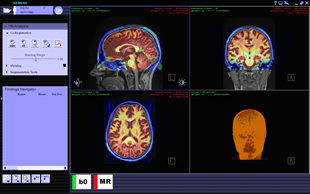
The U.S. Army Medical Research and Materiel Command's Telemedicine and Advanced Technology Research Center is supporting his work, which has two primary thrusts. TATRC has helped Dr. Neiman's team at the ACR connect with all branches of the military to incorporate military healthcare requirements into ongoing industry efforts to standardize medical imaging software. The team is also testing an open-source software framework from Siemens that can efficiently produce applications that are portable and able to run on any system adhering to the emerging application hosting standard.
Notes TATRC Medical Imaging Technologies portfolio manager Dr. Anthony Pacifico, "The technology promises to provide a standardized platform for all to benefit from when studying TBI. It may also have uses in other aspects of radiology."
Neiman's team is developing neuroimaging software through the eXtensible Imaging Platform framework. XIP is an environment for rapidly developing advanced medical imaging applications that are "plug-and-play" and able to work across a wide array of vendor platforms and computing environments. These applications comply with the evolving Digital Imaging and Communications in Medicine draft standard for application hosting (supplement 118).
Says Neiman, "We've used XIP from Siemens to develop software that not only creates higher quality, more accurate 3D images from multiple imaging systems, but that enables a military doctor in Iraq and one at a hospital in the United States to use the same, specialized imaging software to look at the same image and consult over the best treatment. Or a physician following up on an injured warfighter who has been sent home can access recent images with the same software used by the military instead of having to repeat the brain scan."
"Diagnostic techniques are only as good as the visualization tools that support them," he adds. "Gaining one comprehensive image from all available data will aid decision making for radiologists, as they will be able to view regions of interest with greater ease."
The team is currently validating the technology using clinical data.
According to Neiman, positive results will demonstrate that the military research community, using XIP, can rapidly create and deploy portable, DICOM-compliant plug-in applications tailored to the unique imaging needs of injured service members.
"Open-source software is an intriguing, cost-efficient way forward to enhance telemedicine's potential to benefit our warfighters," says TATRC Deputy Director Col. Ron Poropatich. "Software compatibility efforts such as this are also important in the military's move toward electronic health records that physicians can access no matter where the service member is deployed." An official website of the United States government
 ) or https:// means you've safely connected to the .mil website. Share sensitive information only on official, secure websites.


